 Aura: Looksmaxxing
 How to Tell If Your Maxilla Is Recessed
Learn how to tell if your maxilla is recessed using facial landmarks, bite analysis, and self-assessment methods. Practical, anatomy-based guidance.
A recessed maxilla is one of the most commonly discussed structural traits in facial aesthetics, yet most people have no idea whether they have it or not. The maxilla is not a small detail. It is the central foundation of the mid-face, supporting your cheekbones, the base of your nose, your upper teeth, and the positioning of your lips. When it sits too far back relative to the rest of the skull, the effects ripple through nearly every feature on your face.
This guide walks you through the specific visual, functional, and structural signs that indicate maxillary retrusion, how to assess yourself at home with reasonable accuracy, and when it is worth consulting a professional for a more definitive evaluation.
What the Maxilla Actually Is
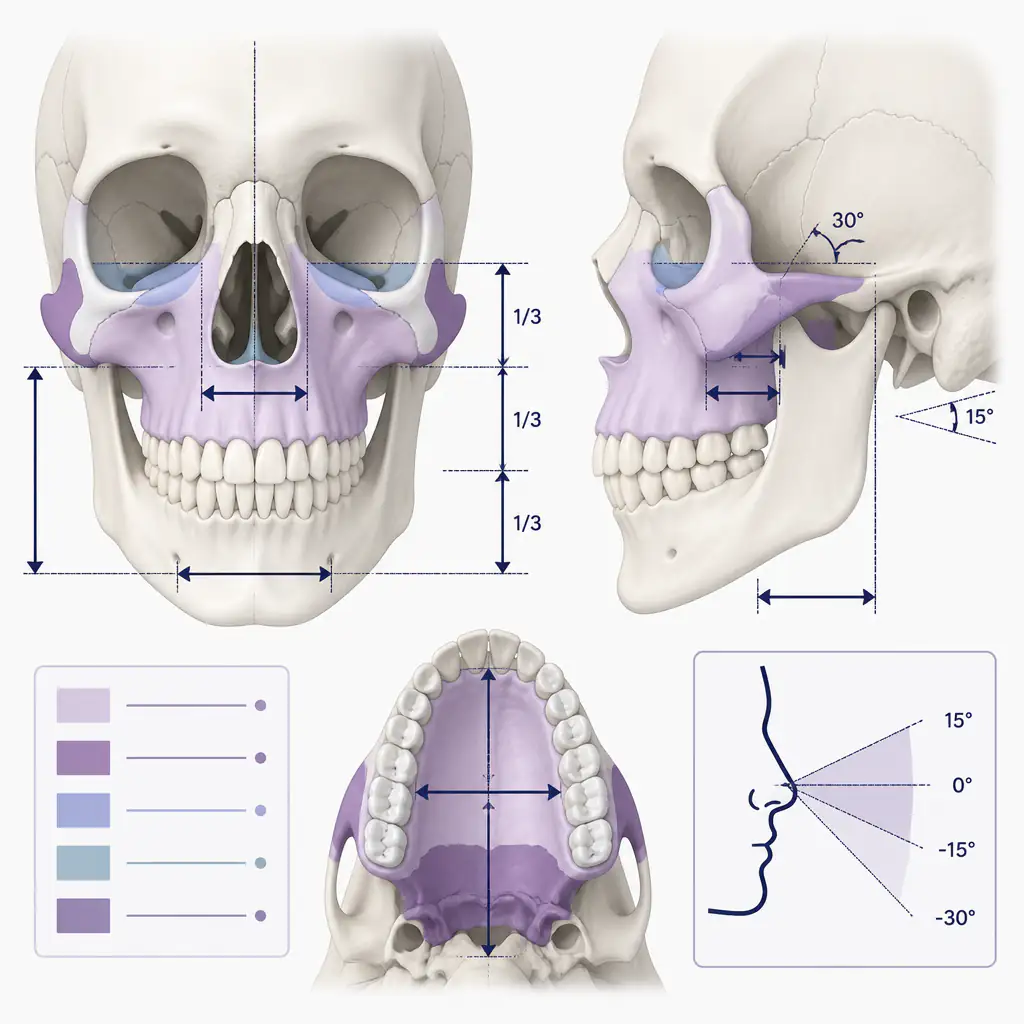
Before assessing yourself, it helps to understand what you are looking at. The maxilla is a paired facial bone that forms the upper jaw, the floor of the nasal cavity, the hard palate, and part of the orbital floor beneath your eyes. In three dimensions, it projects forward and downward from the skull base.
When orthodontists and oral surgeons describe maxillary retrusion or a “recessed maxilla,” they mean the bone sits further back along the horizontal plane (the anteroposterior axis) than is considered ideal. This is distinct from vertical issues like long face syndrome, though the two can overlap.
The formal clinical measurement is called the SNA angle (sella-nasion-A point angle), taken from a lateral cephalometric X-ray. A typical SNA is around 82 degrees. A lower angle suggests the maxilla is retruded. But you do not need an X-ray to notice the surface-level signs.
Common Visual Signs of a Recessed Maxilla
The following visual indicators are not diagnostic on their own, but a combination of several of them is a reasonable signal that the maxilla may be set back.
Flat or Hollow Mid-Face
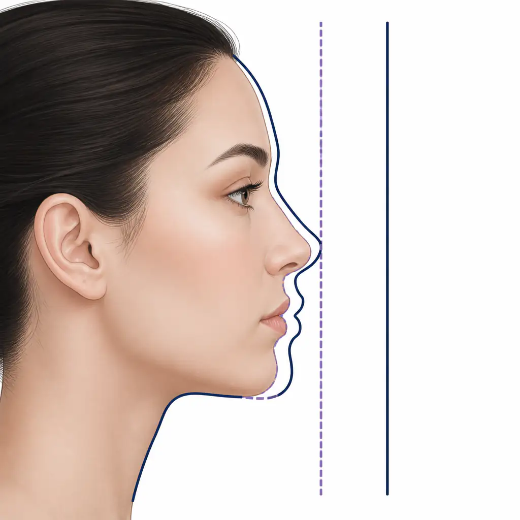
The most obvious sign is a lack of projection in the middle third of the face. When you look at someone from the side, a well-projected maxilla creates a smooth forward curve from the brow down to the lips. A recessed maxilla produces a flatter or even concave profile in that region. The area under the eyes and across the cheeks looks sunken or two-dimensional.
Front-facing, this shows up as under-eye hollows (also called tear troughs or negative orbital vectors) that appear disproportionately deep, even in people who are not underweight.
Lip Posture and Projection
The maxilla is the structural shelf on which the upper lip rests. If the bone is set back, the upper lip tends to follow it inward. This can look like:
- A thin or retruded upper lip at rest
- The lower lip projecting further forward than the upper (called an underbite tendency, even if the teeth do not technically meet in an underbite)
- The nasolabial angle (the angle between the base of the nose and the upper lip) appearing more acute or tucked under
A simple check: look at your side profile in a mirror or photo. Does your upper lip fall behind an imaginary vertical line dropped from the tip of your nose? If it sits notably behind that line while your chin is also behind it, the issue is likely mid-facial rather than just lip volume.
Nose Appearance
The base of the nose attaches directly to the maxilla. When the maxilla lacks forward projection, the nose can appear to droop, rotate downward at the tip, or look overly large relative to the face. Interestingly, some people who believe they need rhinoplasty are actually dealing with a flat mid-face making the nose appear prominent by contrast.
Cheekbone Appearance
High, forward-projecting cheekbones are partly a product of the zygomatic arch, but the maxilla forms the medial (inner) portion of the cheek complex. A recessed maxilla often makes the cheeks appear flat even when the zygomatic arch itself is reasonably wide.
Functional and Dental Signs
Structural issues rarely stay purely cosmetic. The maxilla plays a central role in how your teeth meet and how you breathe, so functional clues can be just as informative as visual ones.
Dental Occlusion
Your bite pattern is one of the most reliable indicators of jaw position. A recessed maxilla is frequently associated with:
- Class III malocclusion: the lower teeth sit in front of the upper teeth when biting down
- Anterior crossbite: some or all of the lower front teeth overlap the upper front teeth
- Underbite: the chin and lower jaw project forward relative to the upper jaw
Note that a Class III bite can also result from a protruding mandible (lower jaw) rather than a recessed maxilla. The distinction matters for treatment. A dentist or orthodontist can differentiate these on examination.
Nasal Breathing Difficulties
The maxilla forms the floor of the nasal cavity. Underdevelopment or retrusion of the maxilla is associated with a narrower palate and reduced nasal airway volume. If you consistently breathe through your mouth, snore, or feel like your nasal passage is narrow even without congestion, it may be worth investigating palate width and maxillary positioning.
Gummy Smile or Excessive Tooth Show
This is a vertical issue that often coexists with horizontal retrusion. If more than 2-3 mm of gum tissue is visible when you smile naturally, or if your upper front teeth are very visible at rest, vertical maxillary excess may be present alongside horizontal retrusion.
The E-Line and Other Profile Checks
A few simple reference lines used by clinicians can be approximated at home using a straight edge or a clear photograph taken from directly to the side.
Ricketts E-Plane
The E-plane (esthetic plane) is a line drawn from the tip of the nose to the tip of the chin. In an ideally balanced adult face:
- The upper lip should sit roughly 4 mm behind the E-plane
- The lower lip should sit roughly 2 mm behind the E-plane
If both lips fall significantly behind this line and the chin is not unusually prominent, the mid-face (including the maxilla) may be underprojected.
The Frankfort Horizontal and Nasion Perpendicular
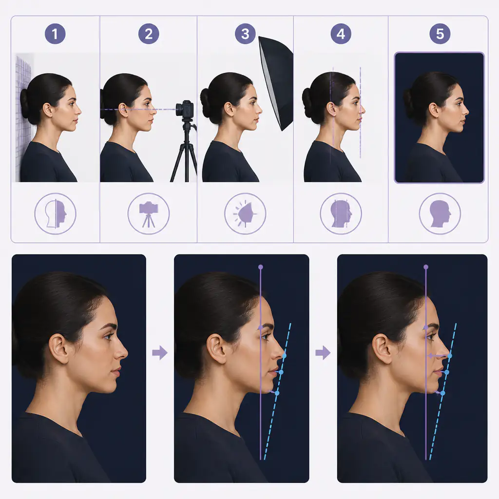
A more precise check involves the nasion perpendicular, a vertical line dropped from the bridge of the nose (nasion). In a well-balanced profile, the upper lip should fall close to this line or slightly behind it. A maxilla that is notably posterior to this reference suggests retrusion.
You can approximate this in a photo: mark the bridge of your nose and draw a vertical line straight down. Check where your upper lip, Cupid’s bow, and philtrum fall relative to that line.
How to Do a Basic Self-Assessment at Home
The following steps will not replace a clinical evaluation, but they can give you a well-informed starting point.
- Take a true lateral photo. Use a camera at eye level, with your head in a neutral position (not tilted up or down). A smartphone propped against a surface works fine. Avoid selfie angles.
- Assess your side profile mid-face. Is there a concavity or flatness from below your eyes to your lips? Does the face look like it caves inward in the middle third?
- Check lip projection. Where does your upper lip fall relative to a vertical line dropped from the tip of your nose?
- Look at your bite in the mirror. Bite down naturally. Do your lower front teeth sit in front of your upper front teeth, or does your lower jaw protrude?
- Check for under-eye hollowness. Look at your face front-on under neutral lighting. Are there pronounced hollows or shadows beneath your eyes that do not change with body weight?
- Note any breathing patterns. Do you default to mouth breathing? Do you have a narrow, high-arched palate?
If multiple items on this list apply to you, it is worth getting a lateral cephalometric X-ray and an evaluation from an orthodontist or oral and maxillofacial surgeon for a definitive answer.
For a structured external baseline, Aura provides AI-based facial analysis that includes mid-face projection scoring, helping you understand how your maxillary area compares to facial harmony benchmarks before committing to any clinical consultation.
What a Recessed Maxilla Does Not Always Look Like
It is worth being clear about what this assessment is not. Not every flat-looking face reflects maxillary retrusion. Factors that can mimic the appearance include:
- Low body fat accentuating bony hollows that would be less visible at a healthy weight
- Age-related volume loss in the cheek fat compartments, unrelated to bone position
- Narrow zygomatic arches with normal maxillary projection
- Purely mandibular prognathism (a forward lower jaw) making the mid-face look relatively set back
This is why a self-assessment based on photos and functional clues is a starting point, not a conclusion.
When to Seek a Professional Evaluation
If your self-assessment raises genuine concerns, the appropriate next steps are:
- Orthodontist consultation: for bite analysis and a referral for cephalometric imaging if needed
- Oral and maxillofacial surgeon (OMFS): for surgical assessment if the structural issue is significant
- Craniofacial orthodontist: for cases involving palate width, airway, and skeletal development in younger patients
Treatment options for a confirmed recessed maxilla range from orthodontic camouflage to palate expansion devices to surgical procedures like Le Fort I osteotomy. The appropriate option depends on the severity of the retrusion, your age, and whether the issue is primarily dental, skeletal, or both.
Always consult a qualified medical or dental professional before considering any intervention for jaw positioning. Self-diagnosis from photos alone is not a substitute for clinical evaluation and imaging.
For those who want a clearer picture of their overall facial structure while gathering information before a professional visit, Aura offers detailed facial analysis including jawline and mid-face metrics that can inform the conversation with your provider.
Summary
A recessed maxilla shows up through a combination of visual, dental, and functional signals: a flat or concave mid-face profile, retruded upper lip, under-eye hollowing, Class III bite tendency, and sometimes chronic mouth breathing. None of these signs is definitive on its own, but multiple signs together provide reasonable grounds for a professional evaluation.
The most actionable thing you can do is take a quality lateral photograph, run through the profile checks described above, and bring your observations to an orthodontist or oral surgeon who can confirm or rule out retrusion with actual imaging. Structural concerns are worth understanding accurately before drawing conclusions or considering any course of action.
Frequently asked questions
Can you tell if your maxilla is recessed just from photos? +
Photos can reveal strong visual indicators like mid-face flatness, retruded lip position, and under-eye hollowing, but they cannot confirm retrusion definitively. A lateral cephalometric X-ray and clinical evaluation are needed to measure the SNA angle and rule out other causes like mandibular prognathism.
Is a recessed maxilla the same as an underbite? +
Not exactly. An underbite means the lower teeth sit in front of the upper teeth when biting, which can result from a recessed maxilla, a protruding lower jaw, or both. Maxillary retrusion is a skeletal position, while an underbite is a dental relationship. They frequently occur together but are assessed separately.
Does a recessed maxilla affect breathing? +
It can. The maxilla forms part of the nasal floor and influences palate width. Underdevelopment or retrusion of the maxilla is associated with a narrower airway, higher rates of mouth breathing, and in some cases sleep-disordered breathing. An ENT or sleep specialist can assess airway involvement.
At what age does maxillary development stop? +
The maxilla continues to grow through adolescence, generally completing development in the mid-to-late teens for females and slightly later for males. Skeletal interventions like palate expansion are more effective before growth plates fuse, which is why early evaluation is often recommended if structural concerns are identified in younger patients.