 Aura: Looksmaxxing
 Bimax Before and After: What to Expect
See what bimax surgery before and after really looks like. Learn what changes, what stays the same, and how to set realistic expectations.
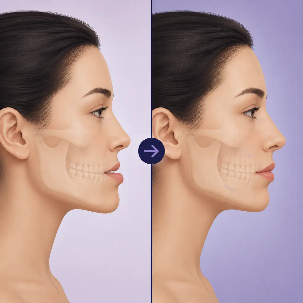
Bimaxillary osteotomy, almost always shortened to “bimax,” is one of the more significant facial surgeries available. It moves both the upper jaw (maxilla) and lower jaw (mandible) simultaneously, which means the changes can be dramatic across the entire midface and lower face. Before and after photos of bimax patients can look striking, sometimes almost like different people, and that naturally draws a lot of curiosity.
But the images circulating online often skip the context: why the surgery was done, what the recovery actually involved, and what a realistic outcome looks like for different starting points. This article breaks down the actual changes you can expect from bimax, what drives good outcomes, and what honest before and after comparisons reveal.
Important: Bimaxillary osteotomy is a major surgical procedure. Always consult a board-certified oral and maxillofacial surgeon or craniofacial surgeon before considering this or any orthognathic surgery.
What Bimax Surgery Actually Does
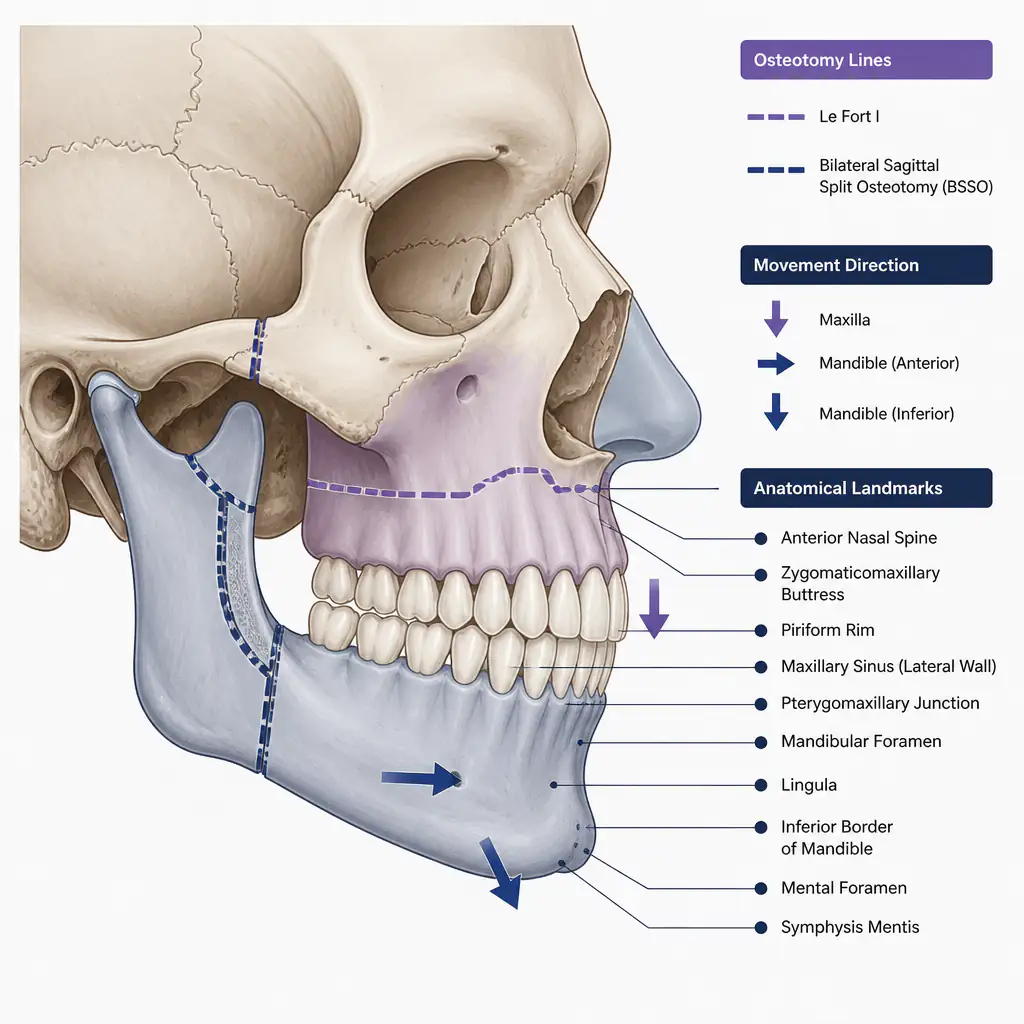
Bimax is a form of orthognathic surgery, meaning it corrects the skeletal structure of the jaws rather than soft tissue alone. The surgeon cuts the maxilla (Le Fort I osteotomy) and the mandible (bilateral sagittal split osteotomy, or BSSO) to reposition both jaws into a new relationship with each other and with the skull base.
The goals are typically:
- Correcting a severe malocclusion (misaligned bite) that cannot be fixed with braces alone
- Improving breathing, chewing, and speech function
- Changing the facial profile as a secondary or co-primary benefit
Because both jaws move together, surgeons can fine-tune the three-dimensional position of the entire lower face. The chin may appear more or less prominent, the lip posture changes, and the nasolabial angle (the angle between the nose and upper lip) often shifts noticeably.
What Changes in Bimax Before and After Photos
Looking at bimax before and after cases, the most consistent changes fall into a few categories.
Jaw Projection and Profile
The most visible change in profile view is jaw projection. If a patient started with a retruded maxilla (sunken midface) and a compensating forward lower jaw, advancement of the maxilla and setback of the mandible can dramatically alter the profile. Conversely, for patients with a skeletal Class II pattern (weak chin, recessed lower jaw), both jaws may be advanced, pushing the entire lower face forward.
In before and after images, this typically shows up as:
- A straighter, more balanced profile line
- Changes in the depth of the philtrum and lip support
- A less convex or less concave facial profile depending on the correction direction
Lip Posture and Support
The maxilla is the structural support for the upper lip. Move the maxilla forward and the upper lip comes with it, appearing fuller and more supported without any soft tissue filler. Move it up (impaction) and lip-to-tooth show at rest improves, reducing a “gummy smile.” These changes are often subtle in frontal photos but obvious in profile.
Chin Projection
Bimax does not directly cut the chin bone (that would be a genioplasty), but repositioning both jaws changes the apparent projection of the chin significantly. Many surgeons combine bimax with genioplasty to fine-tune the chin position after the jaws are set.
Airway and Facial Width
Some patients who undergo maxillary advancement for sleep apnea or airway restriction report a slight broadening of the midface, since the maxilla is being moved forward and its width relationship to the cheekbones subtly changes. This is not always visible but can contribute to a less gaunt appearance.
The Recovery Timeline and How It Affects Results
Bimax before and after comparisons are often taken at wildly different recovery stages, which creates unrealistic impressions online. Understanding the actual timeline helps you read those images more critically.
Week 1 to 4: Significant Swelling
Immediately post-surgery, swelling dominates. The face can look very different from the final result, often puffier and sometimes asymmetric as swelling resolves unevenly. Patients are on a liquid or soft diet, jaw movement is limited, and fatigue is significant.
Month 1 to 3: Early Settling
Most acute swelling resolves in the first six to eight weeks, and patients start to see the emerging result. However, the bones are still consolidating, and the soft tissue is still adapting. Numbness in the lips, chin, and cheeks is common during this period because the inferior alveolar nerve and infraorbital nerve are stretched during the procedure.
Month 3 to 12: Final Result Emerges
Full swelling resolution and nerve recovery can take up to a year. Photos taken at three months will still look slightly different from photos at twelve months. The most accurate “after” photos are taken at least nine to twelve months post-operation.
When you see a bimax before and after comparison with a two-week post-op photo presented as the result, the image is not representative of the final outcome.
What Drives a Good Outcome
Not every bimax result is the same, and the quality of the outcome depends on several factors beyond the surgery itself.
Surgical Planning Quality
Modern orthognathic planning uses 3D CT scans and virtual surgical planning (VSP) software to simulate jaw movements before cutting anything. Surgeons who use precise VSP consistently show better outcomes in published case series. Ask your surgeon specifically about their planning workflow.
Starting Anatomy
Patients with a significant skeletal discrepancy often show the most dramatic change in before and after comparisons, simply because there is more to correct. Someone with a mild Class II malocclusion and adequate lip support will see a more subtle change than someone with a severe Class III (underbite) or a severely retruded maxilla.
Surgeon Experience
Bimax is technically demanding. Volume matters. Surgeons who perform orthognathic procedures regularly, particularly those with fellowship training in oral and maxillofacial surgery or craniofacial surgery, tend to show more consistent results. Reviewing a surgeon’s own before and after gallery (not curated internet photos) is important.
Pre-Surgical Orthodontics
Most bimax patients spend one to two years in braces or aligners before surgery to decompress and align the teeth within each arch. The quality of this pre-surgical orthodontic preparation directly affects how well the jaws can be positioned surgically.
How to Evaluate Bimax Before and After Photos Critically
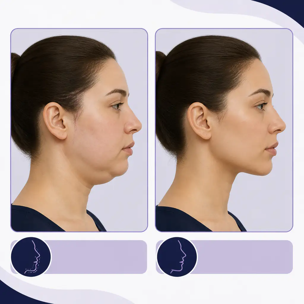
Not all before and after images are created equal. Here is how to read them without being misled.
- Check the time post-op. A result at three months is not a final result. Look for images labeled nine to twelve months or later.
- Check lighting and angle consistency. A slight head tilt or different lighting can make a result look dramatically better or worse than it is. Good comparison images use identical angles and neutral lighting in both shots.
- Look at the occlusion, not just the profile. The primary goal is functional bite correction. A beautiful profile result with a compromised bite is not a successful outcome.
- Consider the starting point. A dramatic transformation is only impressive if the baseline was genuinely severe. Mild corrections will look subtle and that is appropriate.
- Avoid anonymous online galleries. Seek out published case series from peer-reviewed journals or images from credentialed surgeons who are accountable for their outcomes.
If you want an objective baseline of where your own facial structure sits before exploring surgical options, tools like Aura can give you a structured analysis of your jawline, facial proportions, and symmetry, which can be useful context before a surgical consultation.
Bimax vs. Other Jaw Procedures
Bimax is sometimes confused with single-jaw surgeries or other procedures. Here is how they compare.
| Procedure | Jaws Moved | Typical Indication |
|---|---|---|
| Bimax (BSSO + Le Fort I) | Both | Severe skeletal discrepancy, complex malocclusion |
| BSSO alone | Lower only | Mandibular prognathism or retrognathia |
| Le Fort I alone | Upper only | Isolated maxillary issue, gummy smile |
| Genioplasty | Chin only | Chin refinement, often combined with bimax |
| SARPE | Upper (expansion) | Narrow palate, crossbite |
Bimax produces the largest scope of change because both jaws are addressed simultaneously, which also makes it the most involved recovery.
Practical Tips Before Pursuing Bimax
- Start with an orthodontic evaluation. Many patients are referred to an oral surgeon through their orthodontist. This path ensures proper pre-surgical planning.
- Get at least two surgical opinions. Treatment planning for bimax varies, and a second opinion often surfaces different movement options.
- Document your own facial structure carefully. Clear, consistent photos in neutral lighting at multiple angles give you a meaningful baseline for comparison later. Running your photos through Aura can help you understand your current facial metrics objectively before any consultation.
- Understand the functional case first. Insurance coverage for bimax in many regions is tied to documented functional impairment. Having a clear functional diagnosis (bite dysfunction, airway restriction) often determines whether the procedure is covered.
- Budget for the full timeline. Pre-surgical orthodontics, the surgery itself, post-surgical orthodontics, and potential revisions can stretch the process to three or more years total.
Bimax is not a shortcut. The results in genuine before and after comparisons reflect years of planning, orthodontic preparation, surgical execution, and recovery. When those elements align well, the outcomes can meaningfully change both function and facial aesthetics. When they are rushed or poorly planned, the results are inconsistent.
If you are seriously considering this path, work with qualified professionals who can evaluate your specific anatomy, not just photos you found online.
Frequently asked questions
How long does it take to see final bimax results? +
Most surgeons consider the final result visible at nine to twelve months post-operation, once all swelling has resolved and soft tissue has fully adapted. Photos taken earlier than this, especially those taken within the first three months, do not accurately represent the long-term outcome.
Does bimax change the shape of your nose? +
Moving the maxilla forward or upward changes the nasolabial angle, which can make the nose appear slightly rotated or altered in profile. Some patients notice a widening of the nasal base after maxillary advancement. Surgeons may place a cinch suture to manage this, and some patients opt for rhinoplasty separately to address residual nasal changes.
Is bimax surgery purely cosmetic or is it functional? +
Bimax is primarily an orthognathic (functional) surgery used to correct skeletal malocclusions that cannot be resolved with orthodontics alone. Functional goals include correcting the bite, improving chewing efficiency, and in some cases improving the airway. Aesthetic improvement is often a meaningful secondary benefit, but the procedure is not classified as purely cosmetic.
What is the difference between bimax and double jaw surgery? +
They refer to the same procedure. "Double jaw surgery" is the colloquial term for bimaxillary osteotomy, which moves both the upper jaw (maxilla) and lower jaw (mandible). The terms are interchangeable, though surgeons typically use the clinical terminology in formal contexts.