 Aura: Looksmaxxing
 Orbital Box Osteotomy Before and After: What to Expect
Explore orbital box osteotomy before and after outcomes, surgical technique, recovery timeline, and what realistic results actually look like.
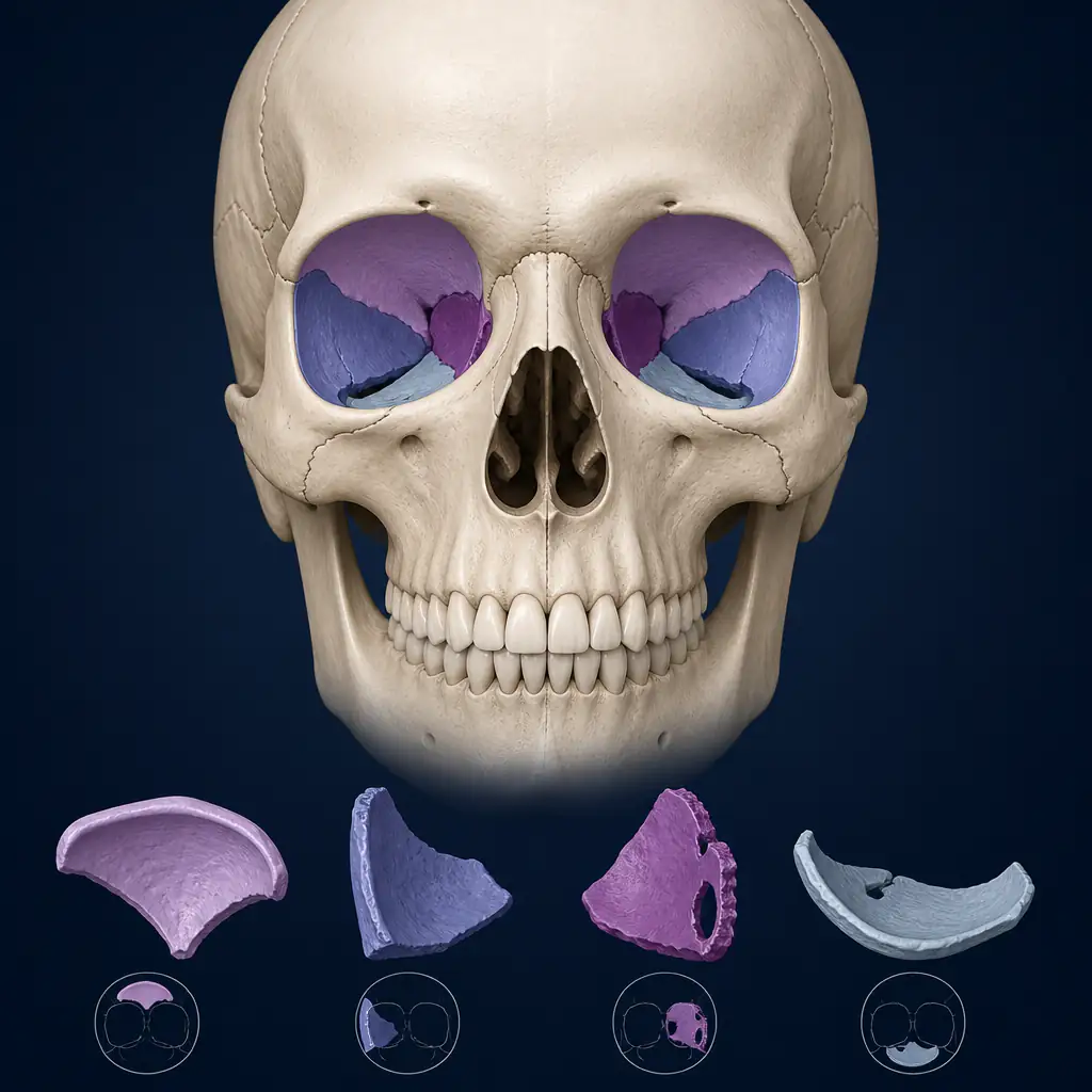
Orbital box osteotomy is one of the more technically demanding procedures in craniofacial surgery. It moves the entire bony orbit, the four-walled box of bone that surrounds the eye, as a single unit. For people seeking significant changes to eye position, orbital rim shape, or facial thirds proportion, this procedure can produce structural changes that softer interventions simply cannot match.
If you are researching orbital box osteotomy before and after results, you are probably trying to answer one of several questions: What exactly changes? How dramatic are the outcomes? What does the recovery involve? This guide works through each of those questions with clinical detail so you can make a genuinely informed decision before speaking with a surgeon.
Important disclaimer: Orbital box osteotomy is a complex craniofacial surgery performed under general anesthesia. It carries serious risks including vision loss, diplopia (double vision), nerve injury, and infection. Talk to a qualified craniofacial or maxillofacial surgeon before considering this procedure. Nothing in this article constitutes medical advice.
What Is an Orbital Box Osteotomy?
The orbit is a bony socket made up of contributions from seven bones: the frontal, zygomatic, maxillary, lacrimal, ethmoid, sphenoid, and palatine bones. In a standard orbital osteotomy, a surgeon cuts through parts of this structure to reposition it. In a box osteotomy specifically, cuts are made on all four walls, allowing the orbit to be moved as a complete, three-dimensional box rather than just repositioning the rim.
This is distinct from simpler orbital rim advancements or zygoma repositioning procedures. Because the whole socket moves together, the eyeball itself moves with it, which changes:
- Globe position (how forward or recessed the eye sits)
- Orbital axis (the angle at which the eye is oriented, relevant for canthal tilt)
- Periorbital bone contour (the visible ridges around the eye)
- Relationship between the eyes and midface
The procedure is used in both reconstructive contexts, such as correcting congenital orbital dystopia (unequal eye heights), and increasingly in aesthetic craniofacial surgery for people seeking structural changes to periorbital appearance.
Who Is This Procedure Designed For?
Candidates tend to fall into a few categories:
Orbital Dystopia Correction
Orbital dystopia means one orbit sits higher, lower, or more lateral than the other. This can be congenital or the result of trauma. A box osteotomy allows surgeons to move one orbit into a position that more closely matches the other, changing both function and appearance.
Aesthetic Orbital Repositioning
Some patients pursue this procedure to address deep-set eyes (enophthalmos), overly prominent eyes (exophthalmos unrelated to thyroid disease), or to modify the horizontal or vertical position of the orbits in relation to the rest of the face. Outcomes in this group depend heavily on the degree of movement attempted and the surgeon’s experience.
Post-Traumatic Reconstruction
After significant orbital fractures or prior surgeries that left the orbit asymmetric, a box osteotomy may be used to re-establish correct three-dimensional position.
For people curious about how their current orbital structure reads aesthetically before considering any intervention, tools like Aura can provide an objective baseline. Aura’s AI-driven facial analysis includes hunter-eye detection, orbital rim assessment, and canthal tilt scoring, which can help frame a conversation with a surgeon around specific measurable targets.
The Surgical Technique: Step by Step
Understanding the technique helps contextualize what before and after comparisons are actually showing.
-
Incision planning. Access is typically gained through a coronal (scalp) incision, sometimes combined with lower eyelid or transconjunctival incisions. This allows broad exposure of the orbital rims and walls with minimal visible scarring.
-
Periorbital dissection. The soft tissues around the orbit are carefully elevated to expose the bony walls. The periorbita (the periosteum lining the orbit) is preserved as much as possible to protect the globe and its supporting structures.
-
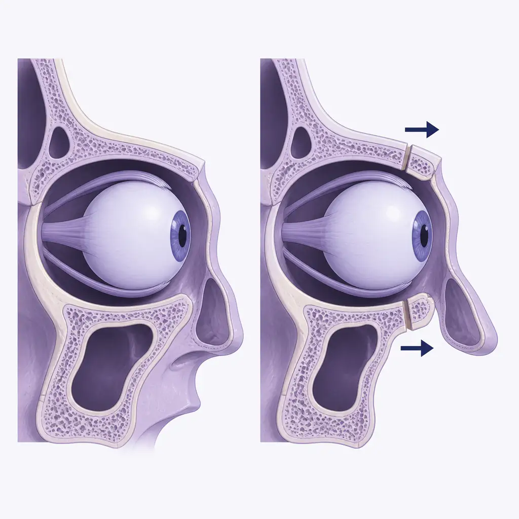
Osteotomies. The surgeon makes precise cuts through the orbital roof (frontal bone), lateral wall (zygoma and greater wing of sphenoid), floor (maxilla), and medial wall (ethmoid and lacrimal bones). The optic canal at the orbital apex is avoided.
-
Mobilization and repositioning. Once the box is free, it is physically moved to the planned coordinates. Movements are typically measured in millimeters. Even a 3 to 5 mm shift can produce a visually significant change.
-
Fixation. The repositioned orbit is secured with titanium plates and screws. Bone gaps created by the movement may be filled with bone grafts or synthetic materials.
-
Closure. Soft tissues are re-draped and incisions are closed in layers.
Total operative time ranges from three to six hours depending on complexity and whether bilateral (both orbits) surgery is planned.
Orbital Box Osteotomy Before and After: What Actually Changes
This is the core of what most people want to understand. Looking at before and after documentation from published case series and board-certified craniofacial surgeons, several consistent patterns emerge.
Globe Position and Eye Shape
Moving the orbit anteriorly (forward) makes the eye appear more prominent and open. Moving it posteriorly gives a more recessed, deep-set appearance. In terms of the PSL-adjacent language many readers in this space are familiar with, anterior movement tends to reduce the appearance of deep-set eyes, while posterior movement can simulate a more recessed orbital look if that is the aesthetic goal.
Canthal Tilt
If the orbital box is rotated slightly during repositioning, the canthal tilt, the angle between the inner and outer corners of the eye, can be meaningfully altered. Surgeons may intentionally plan a degree of rotation to adjust this angle. Before and after photos often show a more positive (upward) or negative (downward) lateral canthal position depending on the rotation applied.
Interorbital Distance
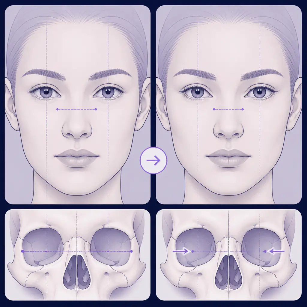
For cases of orbital hypertelorism (eyes set too widely apart), bilateral medial movement of both orbits reduces interorbital distance. This is one of the more visually dramatic changes visible in published before and after series, producing a fundamentally different facial gestalt.
Orbital Rim Contour
The superior orbital rim, which influences brow projection and browbone appearance, may change in prominence. Similarly, the inferior orbital rim’s position affects midface shadow patterns and under-eye contour.
What Does Not Change
The eyelids, canthal ligaments, and soft tissue envelope do not automatically reposition with the bone. Surgeons often perform concurrent soft tissue procedures, such as lateral canthopexy or brow repositioning, to address this. Pure bony movement without soft tissue management can sometimes produce an outcome that looks unusual in photos due to skin laxity or ligament mismatch.
Recovery Timeline and What to Expect
Recovery from orbital box osteotomy is substantial. Setting accurate expectations matters.
Week 1 to 2: Significant periorbital swelling and bruising. Both eyes may swell shut initially. Hospitalization of two to five days is typical. Vision is monitored closely for any signs of optic nerve compromise.
Weeks 3 to 6: Swelling begins to resolve, but the face still looks significantly different from the final outcome. Many patients report difficulty with normal social activities during this window.
Months 3 to 6: The majority of swelling has resolved. Early results become visible. Numbness in the forehead or cheeks from nerve traction begins to improve for most patients, though some cases take longer.
Month 12 and beyond: Final results are typically assessed at one year post-operation. Bone remodeling continues throughout this period. Scar tissue maturation also affects the final soft tissue appearance.
Complications that may affect before and after outcomes include:
- Persistent diplopia (double vision)
- Asymmetry requiring revision
- Numbness from supraorbital or infraorbital nerve involvement
- Orbital volume changes affecting globe position
- Hardware-related issues requiring plate removal
Practical Tips Before Pursuing This Procedure
If you are seriously considering orbital box osteotomy, or even just in the early research phase, a few practical steps can make that process more productive.
Get a detailed facial analysis first. Understanding your current measurements, including orbital rim depth, canthal tilt angle, globe projection, and interorbital distance, gives you a baseline to discuss with surgeons. Aura provides AI-based scoring across several of these dimensions and can help you identify what specifically you are trying to change before committing to any consultation.
Seek craniofacial surgeons specifically. Not all plastic surgeons perform orbital box osteotomies. Look for surgeons with explicit training in craniofacial surgery (often distinguished by fellowship training with groups like the American Society of Maxillofacial Surgeons or equivalent bodies) and ask to see their specific portfolio of orbital cases.
Request three-dimensional imaging. CT-based surgical planning with 3D reconstruction is standard for this procedure at reputable centers. If a surgeon is not using this level of planning, that is a meaningful concern.
Understand the revision rate. Studies on aesthetic orbital surgery suggest revision rates in the range of 10 to 20 percent, often for minor asymmetry corrections or hardware removal. Factor this into your timeline and budget planning.
Be specific about your goals. Surgeons work better with precise anatomical targets than with vague aesthetic preferences. Know whether you are primarily trying to change globe projection, canthal tilt, interorbital distance, or orbital rim contour, and be able to articulate why.
Risks and Limitations
Any honest discussion of orbital box osteotomy before and after results has to address the risk profile. This procedure carries uncommon but serious risks that distinguish it from most facial procedures:
- Vision loss: Rare but documented, resulting from optic nerve injury or vascular compromise. Estimated incidence in experienced hands is under one percent, but the consequence is severe.
- Diplopia: Double vision affects a meaningful percentage of patients post-operatively. Most cases resolve within weeks to months, but persistent diplopia requiring prism correction or strabismus surgery does occur.
- Infection: Given the proximity to the brain and sinuses, orbital infections can have serious consequences including meningitis.
- Asymmetry: Even with precise surgical planning, perfect symmetry is not guaranteed. Minor revisions are common.
These risks do not automatically make the procedure inadvisable for appropriate candidates, but they do require that the decision be made carefully, with realistic expectations and a surgeon whose training and case volume justify the risk.
Comparing Orbital Box Osteotomy to Alternative Approaches
| Approach | What It Changes | Invasiveness | Recovery |
|---|---|---|---|
| Orbital box osteotomy | Full 3D orbit position | Very high | 6-12 months full recovery |
| Lateral canthopexy | Canthal angle only | Low-moderate | 2-4 weeks |
| Orbital rim implants | Rim contour, projection | Moderate | 4-8 weeks |
| Brow bone reduction/augmentation | Superior rim shape | Moderate-high | 6-12 weeks |
| Zygoma repositioning | Lateral orbital wall, midface | High | 3-6 months |
For people whose primary concern is canthal tilt, lateral canthopexy alone may achieve a meaningful portion of the aesthetic change with significantly lower risk. For those with true orbital malposition, structural asymmetry, or goals that softer procedures cannot address, box osteotomy may be the only route to the result they want.
FAQ
Frequently asked questions
How much does orbital box osteotomy cost? +
Costs vary widely by country, surgeon experience, and whether the procedure is bilateral or unilateral. In the United States, all-in costs including anesthesia and facility fees typically range from $30,000 to $80,000 or more for aesthetic cases. Insurance may cover reconstructive cases involving orbital dystopia or post-traumatic deformity.
How long does it take to see final orbital box osteotomy results? +
Most surgeons evaluate final outcomes at 12 months post-operation, as bone remodeling and soft tissue settling continue throughout that period. Early results at three to six months may still show residual swelling or minor asymmetries that improve further over time.
Can orbital box osteotomy change canthal tilt? +
Yes, intentional rotation of the orbital box during repositioning can alter the canthal tilt angle. However, achieving predictable canthal tilt changes often requires concurrent soft tissue procedures such as lateral canthopexy, since the eyelids and canthal ligaments do not automatically reposition with the bone.
Is orbital box osteotomy reversible? +
In practical terms, no. Once osteotomies are made and the orbit is repositioned with titanium fixation, reversing the procedure would require a second major surgery with its own risks and no guarantee of returning to the original anatomy. This is one reason thorough pre-surgical planning and realistic expectations are essential.