 Aura: Looksmaxxing
 Downturned Medial Canthus: What It Means & Options
Learn what a downturned medial canthus is, how it shapes your eye appearance, and what surgical and non-surgical options may help.
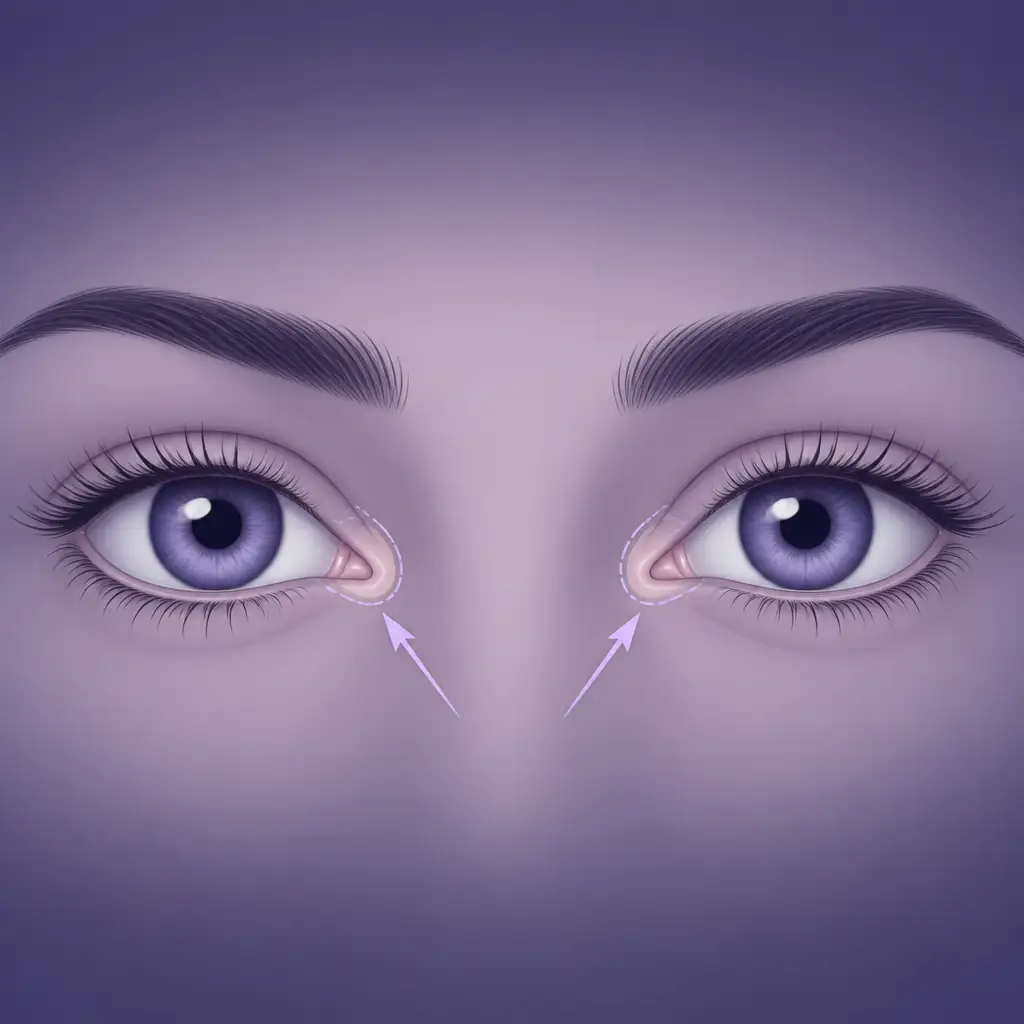
What Is the Medial Canthus?
The medial canthus is the inner corner of the eye, where the upper and lower eyelids meet closest to the nose. It sits at the nasal side of the palpebral fissure (the opening between the eyelids) and is anchored to the bone by a small but structurally important ligament called the medial canthal tendon.
Unlike the lateral canthus (the outer corner), which draws most of the attention in discussions about eye tilt and hunter eyes, the medial canthus has its own distinct influence on how eyes are perceived. Its shape, position, and vertical angle all contribute to the overall character of the eye region.
When people refer to a downturned medial canthus, they mean the inner corner of the eye sits lower than expected relative to the lateral corner, or that the canthus itself angles downward in a way that gives the eye a specific, sometimes described as softer or more melancholic, appearance.
Anatomy Behind the Angle
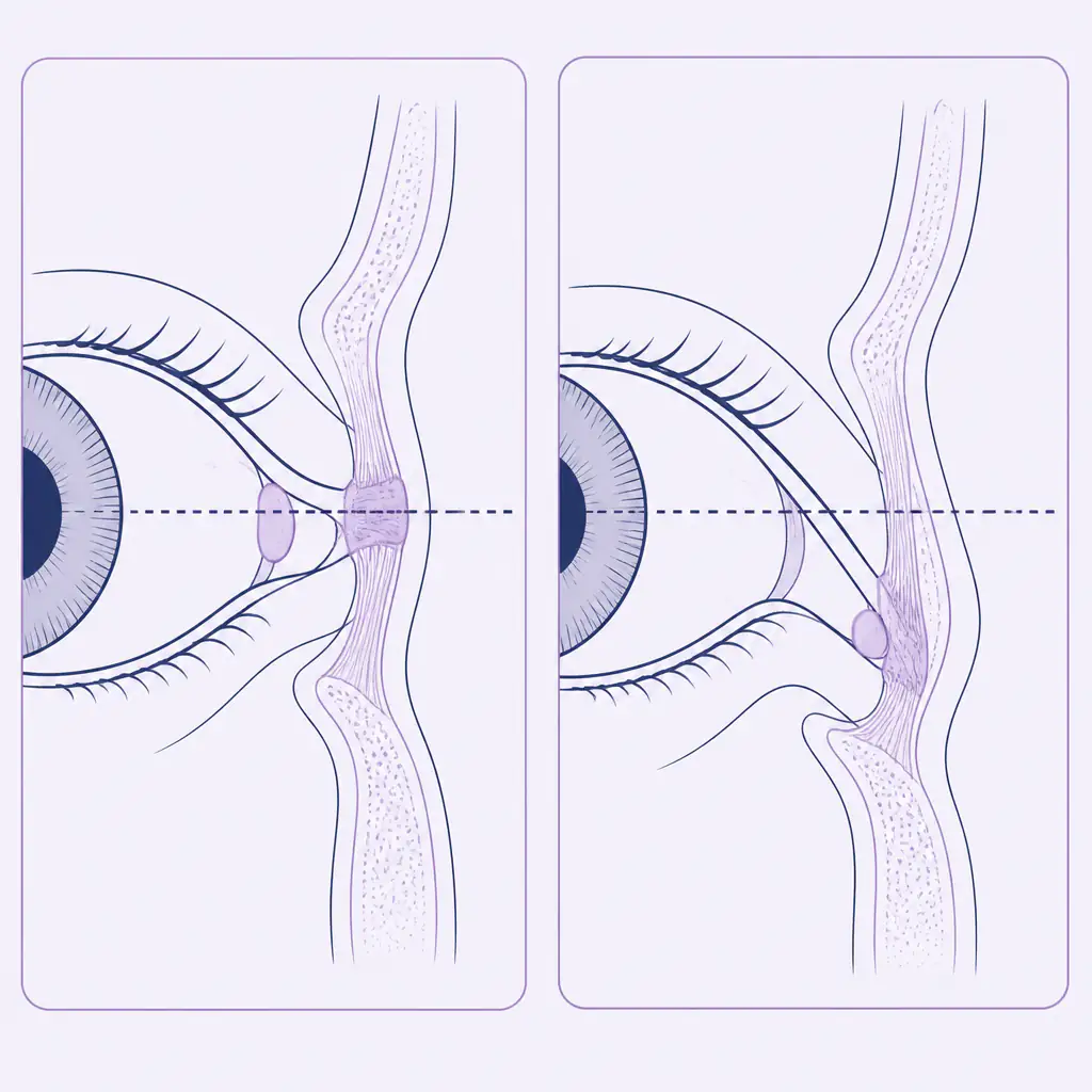
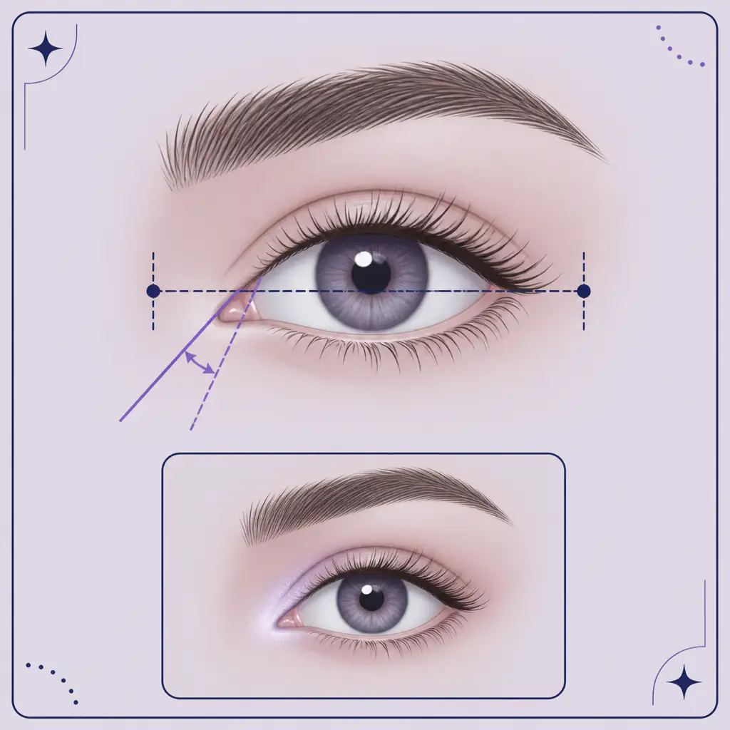
To understand what “downturned” actually means here, it helps to think about the intercanthal axis, an imaginary line drawn between the medial and lateral canthi of the same eye.
- In a neutral or slightly positive canthal tilt, the lateral canthus sits at the same height or slightly above the medial canthus. This is generally associated with a sharper, more alert eye appearance.
- In a negative canthal tilt, the lateral canthus drops below the medial canthus, giving a drooping effect at the outer corner.
- A downturned medial canthus is a slightly different concept: the inner corner itself is pulled or angled downward, which can create the illusion of a teardrop-shaped inner corner, excessive medial scleral show (visible white at the inner eye), or a generally tired appearance even when the lateral tilt is neutral.
The medial canthal tendon attaches to the anterior and posterior lacrimal crests on the medial orbital wall. If this tendon is lax, if the surrounding soft tissue is ptotic (drooping), or if the anatomical position of the inner corner is simply set lower due to genetics, the result is a canthus that sits or points downward.
The lacrimal caruncle, the small pink tissue mass visible at the inner corner, often becomes more prominent or elongated when the medial canthus is downturned, which further changes the perceived shape of the eye.
How a Downturned Medial Canthus Affects Appearance
Aesthetics around the eye are highly sensitive to small changes in geometry. A downturned medial canthus can produce several visual effects:
- Increased medial scleral show. When the inner corner angles down, more of the white (sclera) at the inner eye becomes exposed. This can create a sad or tired look.
- Apparent eye size and shape change. The horizontal axis of the eye shifts, which may make the eye appear rounder or shorter in the horizontal direction.
- Altered perception of facial harmony. The inner canthi are reference points the brain uses when judging symmetry. A downward position on one or both sides can subtly affect how balanced a face looks.
- Interaction with the nose bridge. Because the medial canthus sits close to the nasal root, its angle interacts visually with the width of the nose bridge. A downturned inner corner can emphasize or minimize intercanthal distance (the gap between the two inner corners).
If you want an objective look at how your inner canthal region scores relative to classical proportions, Aura analyzes eye geometry including canthal tilt and gives you a breakdown of what your specific measurements mean for your overall facial rating.
Causes: Why Does This Happen?
A downturned medial canthus can be present from birth or develop over time. The main contributing factors include:
Genetic and Structural Factors
- Inherited bone structure. The shape of the medial orbital wall and the position of the lacrimal crests are determined by genetics. Some ethnic groups have anatomically lower medial canthi as a population-level trait.
- Epicanthal folds. A skin fold covering part of the inner corner (common in East Asian, Central Asian, and some other populations) can visually mimic or physically cause a downward angle at the medial canthus.
- Medial canthal tendon laxity. Some people are born with a slightly looser tendon, which allows the inner corner to drift downward.
Age-Related Changes
As soft tissue volume decreases and ligamentous support loosens with age, the medial canthus can descend. This is part of the broader periorbital aging process that also affects the lower eyelid and tear trough area. Research suggests that medial canthal descent contributes meaningfully to the perception of an aging eye.
Trauma or Previous Surgery
Injury to the medial orbital area or prior eyelid surgery can disrupt the canthal tendon attachment. This is a recognized complication in some rhinoplasty (nose job) procedures that involve the nasal bones close to the canthal anchor points.
Surgical Correction: Medial Canthoplasty
The primary surgical approach for a downturned or malpositioned medial canthus is medial canthoplasty, sometimes combined with epicanthoplasty when an epicanthal fold is involved.
What Medial Canthoplasty Involves
The procedure repositions or tightens the medial canthal tendon to adjust the vertical position and angle of the inner corner. Depending on the goal, surgeons may:
- Tighten the medial canthal tendon by shortening or re-anchoring it to the periosteum (the fibrous layer over bone) at the medial orbital wall.
- Remove or redistribute tissue at the inner corner to change its shape.
- Combine with epicanthoplasty to remove or reposition the epicanthal fold, which often sits directly over the medial canthus and contributes to the downturned appearance.
Recovery typically involves bruising and swelling for one to three weeks, with final results visible after several months as scar tissue softens.
Epicanthoplasty as a Related Procedure
When an epicanthal fold is the primary driver of the appearance, epicanthoplasty alone may be sufficient. This procedure modifies or removes the fold, revealing more of the inner corner and potentially lifting its apparent position. It is one of the more commonly performed aesthetic eye procedures in East and Southeast Asian cosmetic surgery practices.
Important disclaimer: Both medial canthoplasty and epicanthoplasty are surgical procedures with real risks including scarring, asymmetry, dry eye, and damage to the lacrimal drainage system. Talk to a qualified, board-certified oculoplastic or plastic surgeon before considering either procedure. The information here is educational only.
Non-Surgical Approaches and Their Limitations
For people who want to address the appearance without surgery, options are limited but not zero.
Makeup Techniques
Makeup cannot change bone or tendon position, but it can alter visual perception:
- Inner corner highlighting with a light, shimmery product draws the eye upward and inward, creating an illusion of a higher, more pointed inner corner.
- Avoiding dark shadow at the inner corner prevents the area from appearing heavier or more depressed.
- Eyeliner placement along the upper lash line that angles slightly upward from the inner corner can offset the visual effect of a downturned medial canthus.
These are perceptual adjustments, not corrections. They work well for daily appearance management but do not address underlying anatomy.
Filler Considerations
Some practitioners use small amounts of hyaluronic acid filler near the nasal root or tear trough to change how the medial canthal area reads visually. This is an off-label, high-risk injection zone given the proximity to the angular artery and the ophthalmic circulation. Filler in this region carries a non-trivial risk of vascular occlusion and vision complications. It should only be approached by highly experienced injectors, if at all.
Assessing Your Own Medial Canthus
Before exploring any intervention, it is worth getting a clear, objective read on your actual anatomy rather than relying on a single mirror selfie, which distorts facial geometry through lens effects and lighting.
A few practical ways to assess your medial canthus:
- Use a high-quality front-facing photo taken at eye level in neutral light (not from below, which exaggerates the inner corner position).
- Draw or visualize the intercanthal axis. On the same eye, is the inner corner higher, lower, or level with the outer corner?
- Check for epicanthal fold presence. Does skin cover any part of your inner corner? This changes how the canthus angle is measured.
- Assess symmetry between both eyes. Asymmetric medial canthus position is common and may be more noticeable than bilateral downturning.
For a structured analysis, Aura uses AI to measure canthal tilt, eye spacing, and related proportions from a photo and gives you a personalized breakdown, which can be a useful starting point before any conversation with a surgeon or aesthetician.
Practical Tips for Managing the Appearance
If surgery is not on the table and you want to make the most of your current anatomy:
- Control lighting in photos. Overhead lighting casts shadows below the inner corner and exaggerates a downturned appearance. Soft, diffused frontal light is more flattering.
- Work on periorbital skin quality. Puffiness and dark circles in the tear trough area visually compound the effect of a low inner corner. Staying hydrated, reducing sodium, and getting adequate sleep can reduce soft tissue edema around the eye.
- Focus on overall eye support. Strong brow position lifts the entire upper lid complex, which indirectly takes some visual weight off the medial canthal area.
- Consider targeted skincare. Retinoids and peptide-based eye creams may improve skin laxity around the eye over time, though results are modest and gradual.
FAQ
What is a downturned medial canthus?
A downturned medial canthus refers to the inner corner of the eye being angled or positioned lower than typical, either structurally due to genetics or soft tissue, or visually due to the presence of an epicanthal fold. It can affect perceived eye shape and give the eye region a tired or drooping quality.
Is a downturned medial canthus the same as negative canthal tilt?
Not exactly. Negative canthal tilt describes the lateral (outer) corner sitting lower than the medial (inner) corner. A downturned medial canthus describes the inner corner itself being angled or displaced downward, which is a distinct anatomical finding, though both can occur together.
Can medial canthoplasty fix a downturned inner corner?
For appropriate candidates, medial canthoplasty may reposition the inner corner by tightening or re-anchoring the medial canthal tendon. Results depend heavily on the underlying cause and the surgeon’s technique. Talk to a qualified oculoplastic or plastic surgeon to understand whether the procedure is appropriate for your anatomy.
Are there risks to epicanthoplasty for medial canthus correction?
Yes. Epicanthoplasty carries risks including visible scarring at the inner corner (a notoriously difficult area to scar discreetly), asymmetry, over-correction, and recurrence of the fold. Choosing an experienced surgeon who has performed many of these procedures significantly reduces but does not eliminate these risks.
Frequently asked questions
What is a downturned medial canthus? +
A downturned medial canthus refers to the inner corner of the eye being angled or positioned lower than typical, either structurally due to genetics or soft tissue laxity, or visually due to an epicanthal fold. It can affect perceived eye shape and give the eye region a tired or drooping quality.
Is a downturned medial canthus the same as negative canthal tilt? +
Not exactly. Negative canthal tilt describes the lateral (outer) corner sitting lower than the medial (inner) corner. A downturned medial canthus refers to the inner corner itself being angled or displaced downward, which is a distinct anatomical finding, though both conditions can occur together.
Can medial canthoplasty fix a downturned inner corner? +
For appropriate candidates, medial canthoplasty may reposition the inner corner by tightening or re-anchoring the medial canthal tendon. Results depend heavily on the underlying cause and the surgeon's technique. Talk to a qualified oculoplastic or plastic surgeon to determine whether the procedure suits your anatomy.
Are there risks to epicanthoplasty for medial canthus correction? +
Yes. Epicanthoplasty carries risks including visible scarring at the inner corner, asymmetry, over-correction, and fold recurrence. Choosing an experienced surgeon with a strong track record in this specific procedure reduces but does not eliminate these risks.