 Aura: Looksmaxxing
 How to Fix a Recessed Maxilla: Options That Work
Learn how to fix a recessed maxilla with surgical, orthodontic, and lifestyle options. Practical guidance on what actually moves the needle.
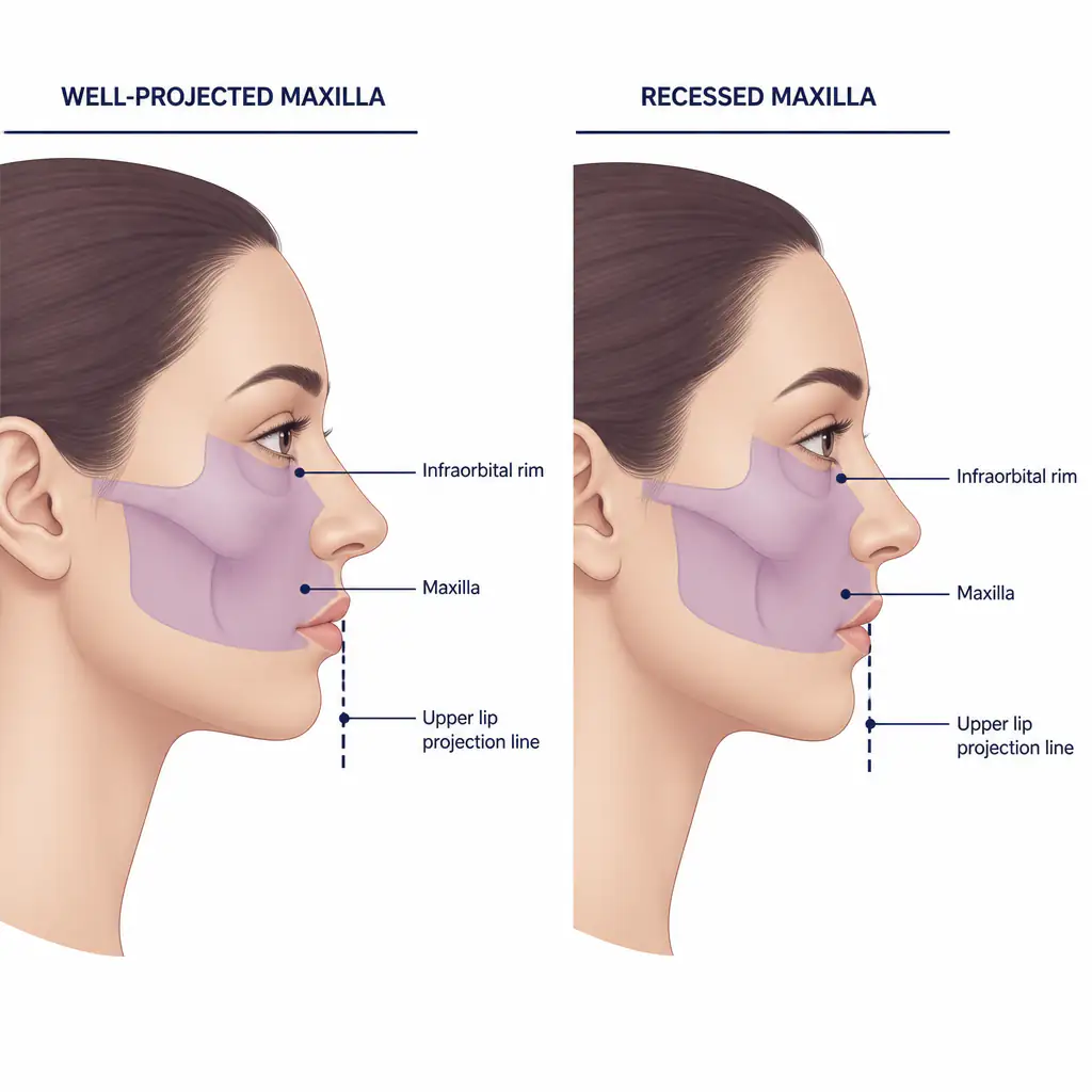
A recessed maxilla is one of the more impactful structural issues affecting facial appearance, yet it often goes undiagnosed or gets blamed on the nose, the chin, or general genetics. If you have been told your midface looks flat, your upper lip lacks projection, or your under-eye area appears hollow, the maxilla is frequently the root cause.
This guide breaks down what a recessed maxilla actually is, why it matters, and what your real options are for addressing it, from non-invasive habits to surgical correction.
What Is a Recessed Maxilla?
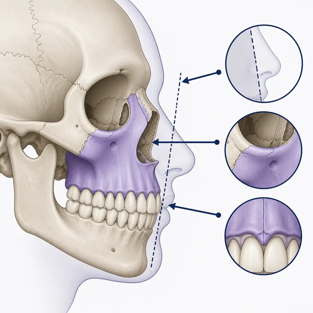
The maxilla is the paired bone that forms most of your midface. It holds your upper teeth, defines the width and projection of your cheekbones, supports the base of your nose, and directly influences the position of your upper lip.
When the maxilla is retrognathic (set back relative to the skull base and lower face), several features tend to cluster together:
- A flat or concave midface profile
- Upper lip that appears thin or retracted
- Increased nasolabial angle (the nose appears to turn upward)
- Hollow or sunken under-eye area (infraorbital rim recession)
- A chin that looks relatively prominent even if it is average in size
- Possible airway narrowing and mouth breathing tendency
Maxillary recession can be skeletal (the bone itself is underdeveloped or set back), dental (upper teeth are tipped inward), or a combination of both. Distinguishing between these categories matters a lot because the appropriate interventions differ significantly.
Why the Maxilla Matters Aesthetically
The midface is the visual anchor of the face. Research in facial attractiveness consistently points to forward midface projection as a marker of strong bone structure. A well-positioned maxilla supports:
- Positive orbital tilt in the under-eye area
- Full upper lip projection without needing fillers
- Defined cheekbone appearance without being overly wide
- Balanced profile where the forehead, midface, and chin sit on a relatively vertical plane
When the maxilla recedes, the nose and chin can appear disproportionate even when they are objectively normal. This is why some people pursue rhinoplasty or chin work and still feel dissatisfied afterward: the underlying structural issue was never addressed.
Before committing to any intervention, getting a clear picture of your current facial structure is worth doing. Aura can give you an AI-based facial analysis, including midface assessment, so you have an objective baseline rather than guessing.
Assessing Your Maxilla: What to Look For
You do not need a surgeon to do an initial self-assessment. Here are some practical ways to evaluate your midface position:
The Profile Test
Stand sideways in front of a mirror or take a neutral lateral photo. Draw an imaginary vertical line down from the most forward point of your forehead (just above the brow). A well-projected maxilla places the upper lip close to or slightly behind this line. If your upper lip falls significantly behind it, midface recession is likely.
The Ricketts E-Plane
The E-plane (esthetic plane) is a line drawn from the tip of the nose to the tip of the chin. In an idealized profile, the upper lip sits slightly behind this line and the lower lip touches it. If your upper lip is noticeably far behind the E-plane, maxillary underprojection may be a factor.
Infraorbital Rim Check
Look straight into a mirror. Place a finger on the bone just below your eye (infraorbital rim). If the rim sits behind the front of your eyeball rather than even with it or slightly in front, you likely have some degree of infraorbital rim recession, which often accompanies maxillary recession.
Dental Relationship
A retrognathic maxilla frequently causes a Class III dental relationship (lower teeth ahead of upper teeth) or contributes to an underbite. Not always, but dental occlusion is a useful data point.
For a deeper structural read, a CBCT (cone beam computed tomography) scan or lateral cephalogram ordered by an orthodontist or oral and maxillofacial surgeon gives precise measurements.
Non-Surgical Approaches
It is important to set realistic expectations here. The maxilla is a bone. Non-surgical methods cannot reposition a fully developed bone the way surgery can. What they can do is optimize posture, oral function, and soft tissue support, which creates a measurable but more modest difference.
Mewing and Proper Tongue Posture
Mewing refers to maintaining the full tongue on the roof of the mouth (the palate) as a resting position. The rationale is that consistent upward and forward tongue pressure may apply gentle mechanical stimulus to the palate and maxilla over time.
The evidence base here is limited, but the practice aligns with well-established orthodontic principles around myofunctional therapy. It is most plausible in adolescents and young adults whose sutures have not fully fused. In adults, effects are likely to be minimal on bone position but may still improve jaw posture, reduce mouth breathing, and support facial muscle tone.
Proper mewing technique:
- Lips gently closed, teeth lightly together or slightly apart
- Entire tongue surface, including the back third, pressed against the palate
- Swallow with the tongue staying on the roof of the mouth
- Maintain this as the default resting position throughout the day
Nasal Breathing
Chronic mouth breathing during growth is associated with longer, narrower faces and less forward maxillary development. In adults, correcting to nasal breathing through myofunctional therapy, treating nasal obstruction, or using nasal strips at night will not move bone, but it can reduce soft tissue changes that worsen with time.
Chewing and Masseter Development
Consistently chewing harder foods (rather than processed, soft diets) and using mastic gum or similar jawline tools may help develop the masseters and temporalis muscles. This does not reposition the maxilla, but stronger facial musculature can slightly improve the visual fullness of the midface.
Posture
Forward head posture pulls the entire craniofacial complex down and back. Correcting neck and spinal posture does not fix skeletal recession, but it can meaningfully change how the face presents at rest and in motion.
Orthodontic and Dental Approaches
For cases where the issue is partly or largely dental (upper teeth tipped inward, a narrow palate, or a functional shift masking true bone position), orthodontic treatment can produce real change.
Palatal Expansion
Rapid palate expansion (RPE) uses a fixed appliance to widen the maxillary arch by gradually separating the mid-palatal suture. In adolescents, this suture is still open, so expansion is straightforward. In adults, the suture is more fused, making conventional RPE less effective and sometimes surgically assisted palate expansion (SARPE) necessary.
Palatal expansion addresses arch width more than forward projection, but widening the arch can improve lip support and reduce the appearance of a flat midface.
Reverse Pull Headgear (Facemask)
A facemask or protraction headgear applies forward traction to the maxilla using hooks on the upper teeth or a palatal expander. This is primarily effective in children (before age 10 to 12) when the midface sutures are still responsive to orthopedic forces. In adults, facemask therapy alone does not move the maxilla meaningfully.
MARPE (Miniscrew-Assisted Rapid Palatal Expansion)
MARPE is a newer orthodontic approach using small titanium screws anchored directly in the palate to apply expansion forces even in adults. Some clinicians report successful suture opening in adults up to their late 20s or early 30s. It is less invasive than surgery and may offer genuine skeletal widening for eligible candidates. Research is still accumulating on long-term outcomes.
Surgical Approaches
For moderate to severe maxillary recession in adults, surgery is the most direct and reliable solution. These are significant procedures, and a qualified oral and maxillofacial surgeon or craniofacial plastic surgeon should be consulted before considering any of them.
Important disclaimer: All surgical procedures carry real risks including infection, nerve damage, relapse, and complications from anesthesia. Talk to a qualified medical professional before considering any surgical intervention.
Le Fort I Osteotomy
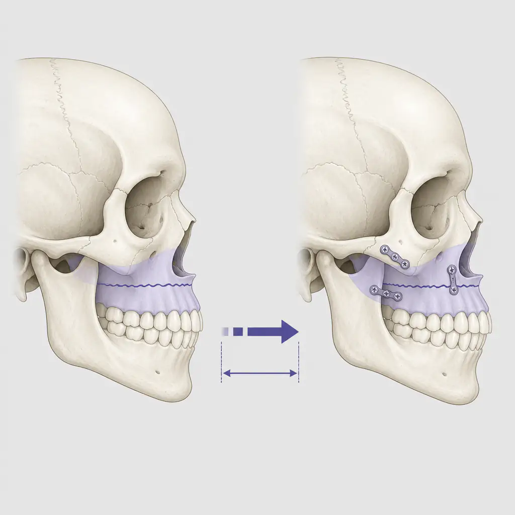
The Le Fort I is the gold-standard surgical procedure for maxillary repositioning. The surgeon cuts the maxilla free from the skull base and physically moves it forward (advancement), upward, or downward depending on the correction needed. It is then fixed in the new position with titanium plates and screws.
Typical outcomes:
- Forward advancement of 4 to 12 millimeters depending on the case
- Improved lip projection and midface fullness
- Correction of Class III dental relationships
- Often combined with genioplasty or mandibular surgery for full facial balance
Recovery involves significant swelling for several weeks, a liquid to soft food diet, and full healing over several months. Results, when the surgical plan is well-designed, are stable and long-lasting.
Bimax Surgery
Bimaxillary surgery combines Le Fort I maxillary advancement with a BSSO (bilateral sagittal split osteotomy) to reposition the lower jaw simultaneously. For cases where both jaws need correction, bimax produces better facial balance than moving one jaw alone.
Implants as an Adjunct
Submalar or midface implants do not fix skeletal recession but can add volume and projection in the infraorbital and cheek region. They are sometimes combined with Le Fort I for patients who need additional soft tissue projection after skeletal repositioning. As a standalone procedure, implants address appearance rather than underlying structure.
Choosing the Right Approach for You
The appropriate intervention depends on several factors:
| Factor | Likely Approach |
|---|---|
| Mild dental tipping only | Orthodontics alone |
| Narrow arch, moderate recession, adolescent | RPE or facemask |
| Narrow arch, adult | MARPE or SARPE |
| Skeletal recession, adult | Le Fort I or bimax surgery |
| Wanting to optimize within current structure | Mewing, posture, myofunctional therapy |
No single approach is right for everyone. Severity, age, budget, risk tolerance, and underlying cause all factor in. A lateral cephalogram and consultation with a board-certified orthodontist and an oral and maxillofacial surgeon together gives you the most complete picture.
If you want to start with a clearer sense of your current facial structure before booking consultations, Aura provides AI-driven facial scoring and midface analysis that can help you frame the right questions for a professional.
Practical Steps to Take Now
- Take a neutral lateral photo in good lighting to assess your profile honestly.
- Start nasal breathing and correct tongue posture regardless of what other steps you pursue. These have no downside and support long-term facial health.
- Book a consultation with an orthodontist who has experience in orthopedic cases and can order a cephalogram.
- If surgery is on your radar, seek out an oral and maxillofacial surgeon who routinely performs orthognathic procedures, not just cosmetic jaw work.
- Do not rush decisions. Orthodontic and surgical plans take months to finalize and years to fully execute. The upside of getting it right is worth the patience.
Recessed maxilla is a well-understood anatomical issue with real, documented solutions. The key is correctly identifying the type and degree of recession, then matching it to the intervention that fits your situation.
Frequently asked questions
Can a recessed maxilla be fixed without surgery? +
In adults, non-surgical methods like mewing, posture correction, and myofunctional therapy may improve facial muscle tone and posture but are unlikely to produce significant skeletal change. Orthodontic options like MARPE may offer modest skeletal expansion for eligible adults. Meaningful correction of moderate to severe maxillary recession in adults generally requires surgery.
How do I know if my maxilla is actually recessed or if it is my nose or chin? +
A lateral cephalometric X-ray taken by an orthodontist or oral surgeon is the most reliable way to assess true maxillary position relative to the skull base. A self-assessment using the Ricketts E-plane or checking the infraorbital rim against the eyeball can give you a rough initial read before booking a professional appointment.
At what age is mewing most effective for the maxilla? +
Tongue posture exercises are most likely to influence bone development during childhood and adolescence, when facial sutures are still open and responsive to mechanical forces. Research suggests that by the mid-20s, the mid-palatal suture is largely fused, significantly limiting any bone-level effect. Adults may still benefit from improved posture and airway function.
What is the recovery like after Le Fort I surgery? +
Most patients experience significant facial swelling for two to four weeks, with dietary restrictions limiting them to liquids and soft foods for six to eight weeks. Most people return to normal activities within four to six weeks, but full bony healing and final aesthetic results can take six to twelve months to fully settle.